
Dr. Arthur Slutsky is one of the world’s most recognized experts in respiratory mechanics, critical care, and mechanical ventilation. His career has bridged biomedical engineering and medicine, shaping the global understanding of positive pressure ventilation (PPV).
- Positions: Professor Emeritus, University of Toronto; Former VP of Research, St. Michael’s Hospital. Chief Scientific Officer, SafeBVM
- Achievements: Hundreds of peer-reviewed articles, major contributions to ventilator-induced lung injury (VILI) prevention, Canadian Medical Hall of Fame inductee (2025).
- Impact: His research has shaped how clinicians worldwide manage acute respiratory failure in ICU and prehospital settings.
Quick Summary / Key Takeaways
If you remember only five things from Dr. Slutsky’s perspective, make it these:
- Mechanical ventilation is indispensable for managing respiratory failure but comes with risks (VILI, pneumonia, diaphragm disuse).
- ARDSNet 2000 (NEJM): low tidal volumes (6 mL/kg PBW) reduced mortality in ARDS.
- Ventilator strategy must be tailored: ARDS (low Vt, higher PEEP), COPD (avoid air-trapping), neuromuscular disease (full support).
- Driving pressure ≤15 cmH₂O is a key target for lung protection in ARDS.
- Automation helps, but waveform interpretation and bedside judgment remain essential.
SECTION 1: Fundamentals & Clinical Indications
FAQ 1: What is mechanical ventilation, and when is it indicated?
Mechanical ventilation uses a machine (a ventilator) to deliver positive pressure to deliver breaths and support or replace spontaneous breathing. It is indicated when patients develop respiratory failure that cannot be managed with supplemental oxygen alone. Common scenarios include ARDS, severe COPD exacerbations, traumatic chest injury, perioperative care during major surgery, drug overdose, and cardiac arrest.
Because it is invasive and carries risks, clinicians weigh the benefits against complications. The timing of intubation has become important — delaying may spare some patients, but waiting too long can worsen outcomes. In the out of hospital setting, it can be used when there is an advanced airway (e.g., ET tube, secure supra-glottic), and is helpful for prolonged ventilation.
Takeaway: While lifesaving, it is invasive and requires careful timing and risk–benefit assessment.
FAQ 2: What are the differences between invasive and non-invasive mechanical ventilation?
Invasive ventilation (endotracheal tube or tracheostomy) ensures control of ventilation and airway protection, but usually needs sedation and carries risks (VAP, airway trauma, delirium).
Non-invasive ventilation (NIV) provides positive pressure via masks or helmets, avoiding an artificial airway. It lowers infection risk and preserves airway reflexes but requires a cooperative patient with intact mental status.
NIV is strongly supported in hypercapnic COPD exacerbations and acute cardiogenic pulmonary edema, whereas severe ARDS generally requires invasive ventilation with protective strategies.
Takeaway: NIV is highly effective in COPD and pulmonary edema, while ARDS usually requires invasive ventilation with protective strategies.
SECTION 2: Risks, Complications & Trade-offs
FAQ 3: What are the most common complications of mechanical ventilation?
Complications are common and clinically significant. Ventilator-induced lung injury (VILI) arises from overdistension (Volutrauma) and can cause Barotrauma (pneumothorax, subcutaneous emphysema). Injury can occur by ventilation at low lung volumes leading to cyclic opening/closing (Atelectrauma). These in turn can lead to Biotrauma, with release of mediators (e.g. IL-6, TNF) into the lung and systemic circulation (PMID: 24283226).
Ventilator-associated pneumonia occurs in ~10–15% of patients, prolonging ICU stays and increasing mortality. Prolonged ventilation promotes diaphragm disuse atrophy and weakness. Sedation needed for ventilation can contribute to delirium and long-term cognitive issues. Prevention bundles and early liberation are vital.
Positive pressure ventilation either with a mechanical ventilator or a BVM can lead to hemodynamic compromise by decreasing venous return and increasing left ventricular afterload.
Takeaway: Prevention bundles, lung-protective strategies, and early liberation are key.
FAQ 4: How does improper ventilator setting selection contribute to patient harm?
Improper settings drive iatrogenic injury. High tidal volumes or plateau pressures cause VILI, while insufficient PEEP promotes atelectrauma. The ARDSNet 2000 NEJM trial showed that tidal volumes of 6 mL/kg PBW reduced mortality versus higher tidal volumes (12 ml/kg PBW), establishing lung-protective ventilation. PEEP must be titrated: too low allows collapse; too high impairs venous return and overdistends alveoli. Rate and I:E ratios affect CO₂ clearance and gas trapping, requiring ongoing reassessment.
Takeaway: Titrate PEEP, tidal volume, and rates carefully — both under- and over-setting can injure.
FAQ 5: Why is weaning from mechanical ventilation so complex?
Weaning requires recovery of respiratory drive, muscle strength, mental status, as well as other critical organs. Barriers include respiratory muscle fatigue (atrophy), excess sedation, delirium, and ICU-acquired weakness. Spontaneous breathing trials and protocols improve success, but failures often reflect cardiac dysfunction, fluid overload, or unresolved illness. Anxiety and agitation can hinder progress, and each failed attempt increases mortality risk.
Takeaway: Structured protocols and spontaneous breathing trials improve success, but each failed attempt worsens outcomes.
SECTION 3: Comparisons, Parameters & Monitoring
FAQ 6: What’s the difference between pressure-controlled and volume-controlled ventilation?
In volume-controlled ventilation, tidal volume is fixed but pressures vary with compliance and resistance of the patient’s respiratory system. In pressure-controlled ventilation, inspiratory pressure is fixed but tidal volume varies with respiratory system mechanics. Volume control ensures predictable minute ventilation; pressure control caps airway pressures and can improve comfort and synchrony. Clinicians switch modes based on goals and changing mechanics.
Takeaway: Volume ensures predictable ventilation; pressure limits airway stress. Clinicians switch based on goals and mechanics.
FAQ 7: How does mechanical ventilation differ between ARDS, COPD, and neuromuscular patients?
Different pathophysiology demands different strategies. In ARDS the lung volume available for ventilation is decreased because of consolidation, atelectasis, and excess fluid. This is often called “baby lung”. Thus, respiratory system compliance is decreased. The goal of ventilation is to deliver adequate gas exchange, but minimize iatrogenic VILI while doing so. So the ventilator strategy uses low tidal volumes, higher PEEP, and often the PaCO2 is allowed to increase. Placing patients in the prone position has also been shown to decreased mortality by limiting VILI (PROSEVA 2013).
A cardinal feature of lung mechanics in patients with COPD is expiratory flow limitation. In these patients excessive minute ventilation can lead to gas-trapping (also called auto-PEEP), which can have a major impact on hemodynamics. Thus, the strategy is to minimize minute ventilation (lower respiratory rates and tidal volumes) to avoid auto-PEEP, even if PaCO₂ increases (controlled hypo-ventilation).
Patients with neuromuscular disease or problems with control of breathing (e.g., opioid overdose) usually have relatively normal lungs but weak respiratory muscles or decreased drive to breathe. In these patients, normal (but relatively low) tidal volumes and respiratory rates can be used to provide full support with while preventing secondary complications.
Takeaway: Ventilation must be disease-specific, not one-size-fits-all.
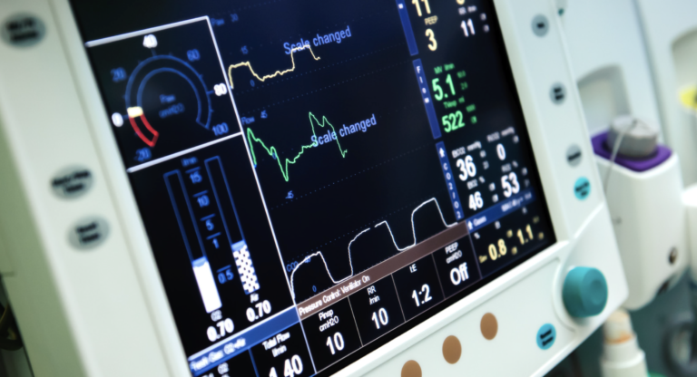
FAQ 8: What role do capnography, plateau pressure, and driving pressure play in ventilator management?
Capnography measures the concentration of CO₂ in exhaled air and displays it as a waveform, providing continuous, non-invasive monitoring of ventilation/perfusion. In the out-of-hospital setting, it is the gold standard for confirming appropriate endotracheal tube placement — a persistent waveform indicates tracheal, not esophageal, intubation. It also helps assess the adequacy of ventilation: rising end-tidal CO₂ (EtCO₂) suggests hypoventilation, while falling values may reflect hyperventilation. Importantly, it is also a measure of perfusion and is decreased during cardiac arrest. During CPR, EtCO₂ levels correlate with cardiac output and can be used to gauge resuscitation quality and identify return of spontaneous circulation (a sudden jump in EtCO₂). In sedated or ventilated patients, capnography provides early warning of airway obstruction, apnea, or disconnection before hypoxemia develops. Overall, it is a vital tool for both safety and physiologic monitoring in emergency and critical care.
Plateau pressure measured during an inspiratory hold in a passive patient can reflect alveolar stress, but it can be misleading in patients who have a stiff chest wall or are obese. Lung protective strategies usually recommend to keep the plateau pressure less than 30 cmH₂O to prevent over-distention of the lungs.
Driving pressure is equal to the plateau pressure minus PEEP. In patients with ARDS it correlates better with mortality that tidal volume or plateau pressure. Targets are often set to keep plateau pressure below 15 cmH₂O to reduce risk.
Takeaway: These measures guide safe ventilation beyond simple Vt and rate.
SECTION 4: Human Factors, Innovation & Clinical Judgment
FAQ 9: Why does mechanical ventilation still require human oversight despite automation?
Patients are variable: compliance, secretions, agitation, pain, and sedation change continuously. Algorithms cannot fully incorporate these nuances easily, especially in a spontaneously breathing patient. Clinicians interpret waveforms, integrate hemodynamics and neurological status, anticipate fatigue, and correct dyssynchrony. However, in trained hands it can be a very useful tool.
Takeaway: Automation assists — but human judgment ensures safety.
FAQ 10: How can providers balance lung protection with gas exchange?
The central challenge is to maintain oxygen delivery while preventing lung injury. In ARDS, prioritize low tidal volumes and limit plateau/driving pressures, accepting permissive hypercapnia if necessary. In COPD, avoiding auto-PEEP and dynamic hyperinflation takes precedence over normalizing CO₂. Adjust PEEP, FiO₂, rate, and Vt dynamically as compliance evolves. ‘Abnormal’ PaCO2 may be the safest plan in some situations, but usually it’s important to maintain adequate oxygenation.
Takeaway: In mechanical ventilation, protect the lungs while ensuring oxygen delivery — use low tidal volumes in ARDS, prevent auto-PEEP in COPD, and accept permissive hypercapnia when safer for the patient.
FAQ 11: What innovations are improving the safety and precision of mechanical ventilation?
Innovations include closed-loop ventilators that adjust PEEP/Vt, AI-assisted protocols, and bedside tools like esophageal manometry and electrical impedance tomography. Earlier in care, smarter bag-valve masks, flow-limiting devices and measuring tidal volumes, respiratory rate, airway pressures and end-tidal CO2 and help improve manual ventilation safety. These tools enhance consistency and precision but still require experienced clinicians to interpret and adjust in real time.
Takeaway: New tools enhance consistency but still need expert oversight.
References
- ARDSNet. N Engl J Med. 2000;342:1301–1308.
- Guérin C, et al. N Engl J Med. 2013;368:2159–2168.
- Wang HE, et al. JAMA. 2018;320(8):769–778.
- Slutsky AS, Ranieri VM. N Engl J Med. 2013;369:2126–36.
- Amato MB, et al. N Engl J Med. 2015;372:747–55.

Haris Shekhani MD, MBID
Haris Shekhani, MD, MBID
- Haris Shekhani MD, MBID
- Haris Shekhani MD, MBID
- Haris Shekhani MD, MBID
- Haris Shekhani MD, MBID
- Haris Shekhani MD, MBID
- Haris Shekhani MD, MBID
- Haris Shekhani MD, MBID
- Haris Shekhani MD, MBID
- Haris Shekhani MD, MBID
- Haris Shekhani MD, MBID
- Haris Shekhani MD, MBID